A comparison of the effects of EDTA on the rate of decrease in organ concentrations of lead vs. organ concentrations of iron was made. Femur lead concentrations of lead-exposed dams were decreased by 77.8% by consumption of the LEAD-X diet, and by 82.3% by consumption of the EDTA diet in comparison with concentrations of dams fed the Basic diet. The corresponding increase in femur iron concentrations was 133.5%, and a decrease in femur iron concentrations was 57.3%.
FIGURE 1 Rat whole blood lead concentrations for dams, day-old pups and week-old pups. Dams were fed diets of Basic, LEAD-X or EDTA diets with or without lead exposure via the drinking water during pregnancy and lactation. Concentrations were significantly affected by LEAD-X(P<0.0001) ,EDTA(P<0.0001)and lead exposure (P<0.0001).
FIGURE 2 Rat brain lead concentration for dams, day-old pups and week-old pups. Dams were fed diets containing Basic, LEAD-X or EDTA diets with or without lead exposure via the drinking water during pregnancy and lactation. Concentrations were significantly affected by LEAD-X(P<0.0001) ,EDTA(P<0.0001)and lead exposure (P<0.0001).
FIGURE 3 Rat femur lead concentration for dams, day-old pups and week-old pups. Dams were fed diets of Basic, LEAD-X or EDTA diets with or without lead exposure via the drinking water during pregnancy and lactation. Concentrations were significantly affected by LEAD-X(P<0.0001) ,EDTA(P<0.0001)and lead exposure (P<0.0002).
FIGURE 4 Rat kidney lead concentration for dams, day-old pups and week-old pups. Dams were fed diets of Basic, LEAD-X or EDTA diets with or without lead exposure via the drinking water during pregnancy and lactation. Concentrations were significantly affected by LEAD-X(P<0.0001) ,EDTA(P<0.0001)and lead exposure (P<0.0001).
FIGURE 5 Rat liver lead concentration for dams, day-old pups and week-old pups. Dams were fed diets of Basic, LEAD-X or EDTA diets with or without lead exposure via the drinking water during pregnancy and lactation. Concentrations were significantly affected by LEAD-X(P=0.0005) ,EDTA(P<0.0001)and lead exposure (P<0.0002).
FIGURE 6 Rat liver iron concentrations for dams, day-old pups and week-old pups. Dams were fed diets containing Basic, LEAD-X or EDTA diets with or without lead exposure via the drinking water during pregnancy and lactation. Iron concentrations were significantly affected by EDTA (P < 0.0001) for the dams, day-old pups and week-old pups. They were significantly affected by lead exposure for the day-old pups (P < 0.0001 ) and week-old pups (P < 0.0001), but not the dams (P > 0.05). There were significant lead, EDTA and LEAD-X interactions.
FIGURE 7 Rat hematocrits for dams, day-old pups and week-old pups. Dams were fed diets containing Basic, LEAD-X or EDTA diets with or without lead exposure via the drinking water during pregnancy and lactation. Hematocrits were significantly affected by dietary EDTA(P<0.0001 ). Lead exposure significantly influenced hematocrits of the day-old pups {P=0.0185}, but not the dams or week-old pups. There were significant lead-EDTA interactions for the day-old and week-old pups , but not for the dams.
FIGURE 8 Rat whole blood hemoglobin concentrations for dams, day-old pups and week-old pups. Dams were fed diets containing Basic, LEAD-X or EDTA diets with or without lead exposure via the drinking water during pregnancy and lactation. Hemoglobin concentrations were significantly affected by LEAD-X(P< 0.0001). Lead exposure significantly influenced hemoglobin concentrations of the dams, day-old pups and week-old pups. There were significant lead-LEAD-X and lead-EDTA interactions.
FIGURE 9 Rat free erythrocyte protoporphyrin concentrations for dams, day-old pups and week-old pups. Dams were fed diets containing Basic, LEAD-X or EDTA diets with or without lead exposure via the drinking water during pregnancy and lactation. Concentrations of FEP were significantly affected by EDTA and by lead exposure.
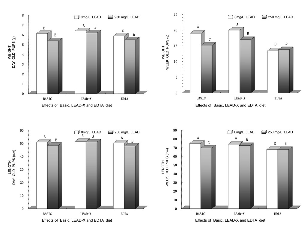
FIGURE I0 Rat fetal pup body weights and lengths for the day-old and week-old pups. Dams were fed diets containing Basic, LEAD-X or EDTA diets with or without lead exposure via the drinking water during pregnancy and lactation. Body weights and lengths of the pups were significantly affected by LEAD-X and by lead exposure.
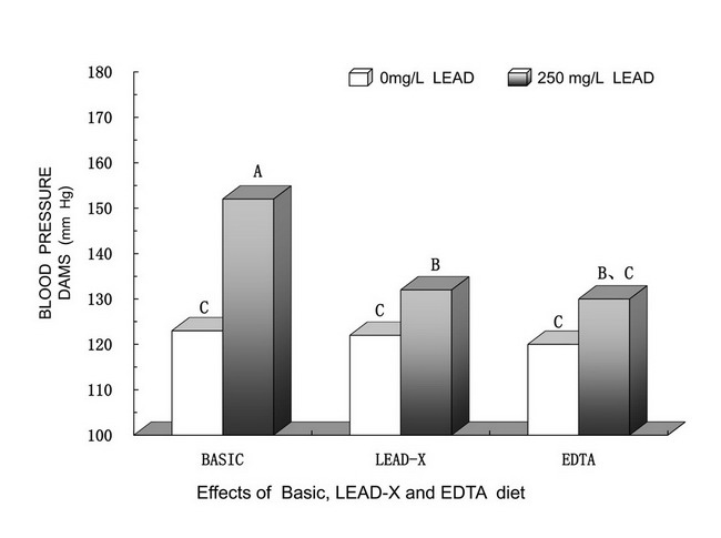
FIGURE 11 Rat systolic blood pressures of dams on d17 of gestation. Dams were fed diets containing Basic, LEAD-X or EDTA diets with or without lead exposure via the drinking water during pregnancy. Blood pressures differed significantly among the treatment groups.
Lead exposure caused modest reductions in hematocrits (Fig.7). However, hematocrits of the dams, day-old pups and week-old pups were reduced considerably by EDTA and basic. A similar pattern was found for the hemoglobin concentrations (Fig.8). Free erythrocyte protopor phyrin concentrations (Fig.9) were increased by lead exposure for dams, day-old pups and week-old pups in the groups fed Basic diet and LEAD-X diet, but not EDTA diet. For the groups fed basic diets, the percentage increase in FEP due to lead exposure was greater in the day-old and week-old pups than in the dams. For the groups fed the LEAD-X diet, FEP concentrations were significantly increased by lead exposure for the dams and day-old pups. The lowest FEP concentrations were found in week-old pups fed the EDTA diet. In fact, consumption of the LEAD-X diet resulted in reduced FEP concentrations exposed to lead. Free erythrocyte protopor phyrin concentrations were increased by EDTA for dams, day-old pups and week-old pups without lead exposure.


